For scientists who study the genetics of hearing and deafness, finding the exact genetic machinery in the inner ear that responds to sound waves and converts them into electrical impulses, the language of the brain, has been something of a holy grail.
The physical basis for hearing and mechanotransduction involves receptor cells deep in the ear that collect vibrations and convert them into electrical signals that run along nerve fibers to areas in the brain where they are interpreted as sound.
What happens in hearing is that mechanical vibration waves traveling from a sound source hit the outer ear, propagate down the ear canal into the middle ear and strike the eardrum. The vibrating eardrum moves a set of delicate bones that communicate the vibrations to a fluid-filled spiral in the inner ear known as the cochlea. .
Inside the cochlea are specialized "hair" cells that have symmetric arrays of extensions known as stereocilia protruding out from their surface. The movement of the fluid inside the cochlea causes the stereocilia to move, and this movement causes ion channels to open. The opening of these channels is a signal monitored by sensory neurons surrounding the hair cells, and when those neurons sense some threshold level of stimulation, they fire, communicating electrical signals to the auditory cortex of the brain.
Scientists at The Scripps Research Institute (TSRI) in La Jolla, CA, have identified a critical component of this ear-to-brain conversion -- a protein called TMHS. This protein is a component of the so-called mechanotransduction channels in the ear, which convert the signals from mechanical sound waves into electrical impulses transmitted to the nervous system. Not only have the scientists finally found a key protein in this process, but the work also suggests a promising new approach toward gene therapy. In the laboratory, the scientists were able to place functional TMHS into the sensory cells for sound perception of newborn deaf mice, restoring their function. In some forms of human deafness, there may be a way to stick these genes back in and fix the cells after birth," TMHS appears to be the direct link between the spring-like mechanism in the inner ear that responds to sound and the machinery that shoots electrical signals to the brain. When the protein is missing in mice, these signals are not sent to their brains and they cannot perceive sound.
Specific genetic forms of this protein have previously been found in people with common inherited forms of deafness, and this discovery would seem to be the first explanation for how these genetic variations account for hearing loss.
TMHS turns out to play a role in a molecular complex called the tip link, which several years ago was discovered to cap the stereocilia protruding out of hair cells. These tip links connect the tops of neighboring stereocilia, bundling them together, and when they are missing the hair cells become splayed apart.
But the tip links do more than just maintain the structure of these bundles. They also house some of the machinery crucial for hearing -- the proteins that physically receive the force of a sound wave and transduce it into electrical impulses by regulating the activity of ion channels. The molecules that form the tip links were identified in the same laboratory previously, but the ion channels and the molecules that connect the tip link to the ion channels remained elusive. For years, scientists have eagerly sought the exact identity of the proteins responsible for this process.
In this study scientists showed that TMHS is one of the lynchpins of this process, where it is a subunit of the ion channel that directly binds to the tip link. When the TMHS protein is missing, otherwise completely normal hair cells lose their ability to send electrical signals.
Journal Reference:
Wei Xiong, Nicolas Grillet, Heather M. Elledge, Thomas F.J. Wagner, Bo Zhao, Kenneth R. Johnson, Piotr Kazmierczak, Ulrich Müller. TMHS Is an Integral Component of the Mechanotransduction Machinery of Cochlear Hair Cells. Cell, 2012; 151 (6): 1283 DOI:10.1016/j.cell.2012.10.041
Scripps Research Institute. "Scientists identify molecules in the ear that convert sound into brain signals." ScienceDaily, 6 Dec. 2012. Web. 20 Feb. 2013.
For the first time, researchers have snapped pictures of mouse inner ear cells using an approach that doesn’t damage tissue or require elaborate dyes. The approach could offer a way to investigate hearing loss and may help guide the placement of cochlear devices or other implants. The small delicate cochlea and associated parts are encased in the densest bone in the body. With standard anatomical imaging techniques such as MRI, the inner ear just looks like a small grey blob. “We can’t biopsy it, we can’t image it, so it’s very difficult to figure out why people are deaf,” said ear surgeon and neuroscientist Konstantina Stankovic of the Massachusetts Eye and Ear Infirmary in Boston. Stankovic and her colleagues took a peek at inner ear cells using an existing technique called two-photon microscopy. Stankovic and her colleagues took a peek at inner ear cells using an existing technique called two-photon microscopy. This approach shoots photons at the target tissue, exciting particular molecules that then emit light. The researchers worked with mice exposed to 160 decibels of sound for two hours. Then they removed the rodents’ inner ears, which includes the spiraled, snail-shaped cochlea and other organs. Instead of cutting into the cochlea, the researchers peered through the “round window” — a middle ear opening covered by a thin membrane that leads to the cochlea.
The approach yielded clear images of rows of the inner ear’s hair cells, tiny hairlike structures that detect sound vibrations, enabling hearing. Unlike in control mice, ears of noise-exposed mice had visible damage: Whole sections of ear cells were wiped out, Stankovic reported February 17 at the annual meeting of the American Association for the Advancement of Science.
She hopes the approach will not only shed light on the various kinds of damage that lead to hearing loss, but that it also will help guide the insertion of implants. In the future, the imaging approach might help guide the placement of an experimental device that extracts energy from the inner ear, acting as a tiny battery. The new device, developed by Stankovic and colleagues, doesn’t generate enough power to run a cochlear implant. It could, however, act as a sensor, monitoring for infections or sensing drug levels.
Such devices might prove very useful for monitoring all sorts of physiological responses, says biomedical engineer Philippe Renaud of Swiss Federal Institute of Technology in Lausanne. They could prove especially useful, he says, in patients for whom devices need to be small, delicate and efficient, such as those undergoing deep brain stimulation.
Citations:
K. M. Stankovic. Treating deafness with better vision: cellular level optical imaging of the inner ear. Annual meeting of the American Association for the Advancement of Science, Boston. Presented February 17, 2013
Imaging technique offers look inside hearing loss
By Rachel Ehrenberg in Science news
Gene therapy is the use of DNS as a pharmaceutical agent to treat disease. It derives its name from the idea that DNA can be used to supplement or alter genes within an individual's cells as a therapy to treat disease. The most common form of gene therapy involves using DNA that encodes a functional, therapeutic gene to replace a mutated gene. Other forms involve directly correcting a mutation, or using DNA that encodes a therapeutic protein drug (rather than a natural human gene) to provide treatment. In gene therapy, DNA that encodes a therapeutic protein is packaged within a "vector", which is used to get the DNA inside cells within the body. Once inside, the DNA becomes expressed by the cell machinery, resulting in the production of therapeutic protein, which in turn treats the patient's disease.
Gene therapy using an Adenovirus vector. A new gene is inserted into a cell using an adenovirus. If the treatment is successful, the new gene will make functional protein to treat a disease.
Gene therapy is still in its infancy, with obvious challenges around targeting damaged cells and creating corrective genes. An equally important challenge, addressed by this research, is finding ways to transport the corrective genes into the cell. This is a problem, because of the poor permeability of cell membranes. There are several barriers to gene delivery, The genetic material must be protected during transit to a cell, it must pass into a cell, it must survive the cell's defense mechanisms, and it must enter into the cell's guarded nucleus.
If all of these barriers can be overcome, gene therapy would be a valuable technique with profound clinical implications. It has the potential to correct a number of human diseases that result from specific genes in a person's DNA makeup not functioning properly - or not at all. Gene therapy would provide a mechanism to replace these specific genes, swapping out the bad for the good. Safety is a primary concern when working with gene therapy. Some of the first attempts at gene therapy used viruses to insert DNA into cells. Viruses can be dangerously toxic, however, and this fact was tragically demonstrated a decade ago when an 18-year-old boy enrolled in a gene therapy study had a massive immune reaction to the viruses used. He died just a few days into the treatment from multiple organ failure, precipitating an immediate halt to the trial.
Several patients in gene therapy clinical trials have developed leukemia as a result of their treatment. The underlying cause of leukemia is thought to be that the viral vectors used to carry the therapeutic gene into cells (gamma-RVs) integrate into the genome of the cells disrupting the natural control of cancer-associated genes (a process known as insertional mutagenesis).
In gene therapy, the viruses are often equipped with additional genes, such as for immune mediators or for proteins inducing programmed cell death. However, these gene products can harm the body if they are released at the wrong moment or at excessive levels. It would be advantageous, if these switches could be turned on and off at a specific time. In order to construct such a switch, the researchers inserted synthetic segments of DNA into the viral genetic material in the direct vicinity of the transferred gene. In the infected cell, this construct is transcribed together with the transferred gene into a single messenger RNA (mRNA) molecule. The switch is operated using an agent which is added to cells infected with the virus. The substance is precisely fitted to bind to the RNA molecule and induces it to cut itself up. Thus, the potentially dangerous protein cannot be produced. The researchers copied this regulation mechanism from bacteria which use RNA switches to regulate production of numerous proteins.
Since then, many alternatives to viruses have emerged for use in gene therapy, including synthetic molecules like "dendrimers," a word that derives from the Greek word for "tree." Similar to trees, dendrimers are branching molecules that are slightly positively charged. This allows them to be loaded with DNA (which is slightly negative charged) for insertion into a cell. Dendrimers seem to offer many advantages over viruses. They may be much less toxic, and they may offer other advantages in terms of cost, ease of production, and the ability to transport very long genes. If they can be designed to efficiently -- and safely -- shuttle genes into human cells, then they may be a more practical solution to gene therapy than viruses. So far, laboratory experiments with different types of dendrimers have shown that they can insert genes into cells, but only with very low efficiency.
Using the principles of evolution and natural selection, that were initially conceived by Charles Darwin, they have now developped an efficient and safe gene delivery approach based on non-viral genetic elements, called transposons. Transposons are mobile DNA elements that can integrate into 'foreign' DNA via a 'cut-and-paste' mechanism. In a way they are natural gene delivery vehicles. The researchers constructed the transposons in such a way that they can carry the therapeutic gene into the target cell DNA. Doing so, they obviate the need to rely on viral vectors
This research describes a model peptide sequence, dubbed GeT (gene transporter), which wraps around genes, transports them through cell membranes and helps their escape from intracellular degradation traps. The process mimics the mechanisms viruses use to infect human cells. GeT was designed to undergo differential membrane-induced folding -- a process whereby the peptide changes its structure in response to only one type of membranes. This enables the peptide, and viruses, to carry genes into the cell. Interestingly, the property also makes it antibacterial and so capable of gene transfer even in bacteria-challenged environments. To prove the concept, the researchers used GeT to transfer a synthetic gene encoding for a green fluorescent protein -- a protein whose fluorescence in cells can be seen and monitored using fluorescence microscopy. The design can serve as a potential template for non-viral delivery systems and specialist treatments of genetic disorders.
To create the new gene therapy vector, Jans and colleagues used pieces of different genes to create a protein called a "modular DNA carrier," which can be produced by bacteria. This protein carries therapeutic DNA and delivers it to a cell's nucleus, where it reprograms a cell to function properly. In the laboratory, these carrier proteins were combined with therapeutic DNA and attached to cell membrane receptors and the nuclear import machinery of target cells. In turn, the packaged DNA moved into the cell through the cytoplasm and into the nucleus.
Genetically-engineered spider-silk proteins represent a versatile and useful new platform polymer for non viral gene delivery. The scientists describe modifying spider silk proteins so that they attach to diseased cells and not healthy cells. They also engineered the spider silk to contain a gene that codes for the protein that makes fireflies glow in order to provide a visual signal (seen using special equipment) that the gene has reached its intended target. In lab studies using mice containing human breast cancer cells, the spider-silk proteins attached to the cancer cells and injected the DNA material into the cells without harming the mice.
For the first time ever, chitosan nanoparticles have been used as a carrier for gene therapy in the ear. Chitosan is produced from shrimp shells. researchers attempted to use chitosan as a carrier to deliver drugs and genes to the inner ear in guinea pigs. Chitosan was able to deliver drugs through the membrane that covers the tiny gap between the middle ear and inner ear. Chitosan was also able to deliver genes to the hair cells. Whether or not the results from guinea pigs can be transferred to human ears remains uncertain. However, chitosan is non-toxic and is not harmful to cells. Chitosan is therefore better than other carriers and has characteristics that mean it could potentially be used with patients. Extremely small nanoparticles in the range of 50-200 nm (nanometres) are formed spontaneously when the positively charged chitosan and negatively charged genes are mixed. Chitosan does a good job packaging up DNA and RNA's relatively large molecules. When the nanoparticles have passed through a membrane, chitosan packages up the gene molecules so they return to their normal size again. Chitosan also creates gaps between cells, which facilitate the absorption of medicine.
Researchers from Johns Hopkins and Northwestern universities have discovered how to control the shape of nanoparticles that move DNA through the body and have shown that the shapes of these carriers may make a big difference in how well they work in treating cancer and other diseases. A major advance in this work is that Mao and his colleagues reported that they were able to "tune" these particles in three shapes, resembling rods, worms and spheres, which mimic the shapes and sizes of viral particles. "We could observe these shapes in the lab, but we did not fully understand why they assumed these shapes and how to control the process well," Mao said. These questions were important because the DNA delivery system he envisions may require specific, uniform shapes. The worm-shaped particles resulted in 1,600 times more gene expression in the liver cells than the other shapes," Mao said. This means that producing nanoparticles in this particular shape could be the more efficient way to deliver gene therapy to these cells. The particle shapes used in this research are formed by packaging the DNA with polymers and exposing them to various dilutions of an organic solvent. DNA's aversion to the solvent, with the help of the team's designed polymer, causes the nanoparticles to contract into a certain shape with a "shield" around the genetic material to protect it from being cleared by immune cells.
Source:
Wikipedia
VIB (the Flanders Institute for Biotechnology) (2009, May 9). Moving Gene Therapy Forward With Mobile DNA. ScienceDaily. Retrieved January 13, 2013, from http://www.sciencedaily.com/releases/2009/05/090503132615.htm
Federation of American Societies for Experimental Biology (2009, September 2). Finding The ZIP-code For Gene Therapy: Scientists Imitate Viruses To Deliver Therapeutic Genes. ScienceDaily. Retrieved January 13, 2013, from http://www.sciencedaily.com/releases/2009/08/090831130749.htm
Helmholtz Association of German Research Centres (2012, August 21). Viruses with integrated gene switch. ScienceDaily. Retrieved January 13, 2013, from http://www.sciencedaily.com/releases/2012/08/120821114738.htm
National Physical Laboratory (2011, August 12). Scientists copy the ways viruses deliver genes.ScienceDaily. Retrieved January 13, 2013, from http://www.sciencedaily.com/releases/2011/08/110811094836.htm
American Institute of Physics (2009, April 30). First Large-scale Computer Simulation Of Gene Therapy. ScienceDaily. Retrieved January 13, 2013, from http://www.sciencedaily.com/releases/2009/04/090429152430.htm
Northwestern University. "Shape matters in DNA nanoparticle therapy: Particles could become a safer, more effective delivery vehicle for gene therapy." ScienceDaily, 12 Oct. 2012. Web. 13 Jan. 2013.
Tinnitus is defined as the perception of sound in the absence of external sound and can manifest itself in variety of ways. The phantom sounds of tinnitus may sound like ringing, clicking or hissing. The disorder is most often caused by damage to the microscopic endings of the hearing nerve in the inner ear, although it can also be attributed to allergies, high or low blood pressure, a tumor, diabetes, thyroid problems, injury to the head or neck, and use of medications such as anti-inflammatories, antibiotics, sedatives, antidepressants, and aspirin.
New research at the University of Michigan Health System suggests over-exposure to noise can actually cause more lasting changes to our auditory circuitry -- changes that may lead to tinnitus, commonly known as ringing in the ears. U-M researchers previously demonstrated that after hearing damage, touch-sensing "somatosensory" nerves in the face and neck can become overactive, seeming to overcompensate for the loss of auditory input in a way the brain interprets -- or "hears" -- as noise that isn't really there. The new study, which appears in the Feb. 1 issue of The Journal of Neuroscience, found that somatosensory neurons maintain a high level of activity following exposure to loud noise, even after hearing itself returns to normal. In normal hearing, a part of the brain called the dorsal cochlear nucleus is the first stop for signals arriving from the ear via the auditory nerve. But it's also a hub where "multitasking" neurons process other sensory signals, such as touch, together with hearing information. During hearing loss, the other sensory signals entering the dorsal cochlear nucleus are amplified, This overcompensation by the somatosensory neurons, which carry information about touch, vibration, skin temperature and pain, is believed to fuel tinnitus in many cases. The involvement of touch sensing (or "somatosensory") nerves in the head and neck explains why many tinnitus sufferers can change the volume and pitch of the sound by clenching their jaw, or moving their head and neck, "This is the first research to show that, in the animals that developed tinnitus after hearing returned to normal, increased excitation from the somatosensory nerves in the head and neck continued.
Prior research has shown that auditory circuits in the brain are more excitable in tinnitus sufferers, but until now it has not been clear whether that is due to hyperactivity of excitatory neural pathways, reduced activity of inhibitory ones, or a bit of both, Thanos Tzounopoulos, Ph.D., assistant professor of otolaryngology and neurobiology, Pitt School of Medicine used a new technique to image auditory circuits using slices of brain tissue in the lab. Dr. Tzounopoulos' team created tinnitus in a mouse model. The scientists then sought to determine what had gone wrong in the balance of excitation and inhibition of the auditory circuits in the affected mice. They established that an imaging technique called flavoprotein autofluorescence (FA) could be used to reveal tinnitus-related hyperactivity in slices of the brain. Experiments were performed in the dorsal cochlear nucleus (DCN), a specialized auditory brain center that is crucial in the triggering of tinnitus. FA imaging showed that the tinnitus group had, as expected, a greater response than the control group to electrical stimulation. Most importantly, despite local stimulation, DCN responses spread farther in the affected mice. Dr. Tzounopoulos' new experimental approach has resolved why tinnitus-affected auditory centers show increased responsiveness. After administering a variety of agents that block specific excitatory and inhibitory receptors and seeing how the brain center responded, his team determined that blocking an inhibitory pathway that produces GABA, an inhibitory neurotransmitter, enhanced the response in the region surrounding the DCN in the control brain slices more so than it did in the tinnitus slices. This means that agents that increase GABA-mediated inhibition might be effective treatments for tinnitus. Dr. Tzounopoulos' team is now trying to identify such drugs.
Because subjective tinnitus is typically localized to the ear with hearing loss, tinnitus was traditionally thought to originate from neural hyperactivity in the damaged ear. However, most studies have found that hearing loss reduces the neural outputs from the damaged cochlea. These negative findings led to the hypothesis that rinnitus arises from aberrant neural activity in the central auditory system. Positron emission tomography imaging studies performed on tinnitus patients that could modulate their tinnitus provide evidence showing that the aberrant neural activity that gives rise to tinnitus resides in the central auditory pathway.
A study conducted at the University of Arkansas for Medical Sciences (UAMS) has shown potential to markedly improve tinnitus. The study aimed at examining the safety and feasibility of using maintenance sessions of low-frequency repetitive transcranial magnetic stimulation (TMS) to reduce tinnitus loudness and prevent its return over time. TMS involved the placement of a coil on the scalp that creates a magnetic field over the brain’s surface. The magnetic field penetrates up to two or three centimeters from the surface of the coil. An electric current is induced by the magnetic field that either activates or inhibits neural activity. The goal of the study was to inhibit excessive neural activity believed to cause tinnitus. They used a PET scan of the patient’s brain to look for excessive neural activity with increased blood flow in the temporal lobe. They then targetted that area with low-frequency TMS to inhibit the neural activity and decrease the tinnitus.
Researchers Dr. Michael Kilgard and Dr. Navzer Engineer from The University of Texas at Dallas and University-affiliated biotechnology firm MicroTransponder report that stimulation of the vagus nerve paired with sounds eliminated tinnitus in rats. The auditory cortex delegates too many neurons to some frequencies. Because there are too many neurons processing the same frequencies, they are firing much stronger than they should be. In addition, the neurons fire in sync with one another and they also fire more frequently when it is quiet. It's these changing brain patterns that produce tinnitus. Brain changes in response to nerve damage or cochlear trauma cause irregular neural activity believed to be responsible for many types of chronic pain and tinnitus. But when they paired tones with brief pulses of vagus nerve stimulation, they eliminated the physiological and behavioral symptoms of tinnitus in noise-exposed rats. The researchers are, in essence, retraining the brain to ignore the nerve signals that simulate ringing. This minimally invasive method of generating neural plasticity is used to precisely manipulate brain circuits, which cannot be achieved with drugs, "Pairing sounds with VNS provides that precision by rewiring damaged circuits and reversing the abnormal activity that generates the phantom sound.
Sound masking therapy, a common component of tinnitus treatment, is of uncertain benefit when used on its own, a new evidence review finds. Sound therapy relies on distraction, with an additional noise -- often called white noise -- reducing the contrast between the patient's tinnitus signal and the background noise. This reduces the person's perception of the phantom sounds and the distress they cause. People receive sound therapy in several ways: by devices worn in or on the ear, by headphones connected to an MP3-like device or with an under-the-pillow speaker for night use. the weight of evidence does not support sound therapy as a primary treatment. Sound therapy is useful only when combined with a counseling component to complete a therapeutic process."
Shaowen Bao's experiments with induced hearing loss in rats explain why the neurons in the auditory cortex generate these phantom perceptions. They showed that neurons that have lost sensory input from the ear become more excitable and fire spontaneously, primarily because these nerves have "homeostatic" mechanisms to keep their overall firing rate constant no matter what. With the loss of hearing, you have phantom sounds. Tinnitus resembles phantom limb pain experienced by many amputees, One treatment strategy, then, is to retrain patients so that these brain cells get new input, which should reduce spontaneous firing. This can be done by enhancing the response to frequencies near the lost frequencies. They changed their (brain training) strategy from one where they completely avoided the tinnitus domain to one where they directly engage it and try to redifferentiate or reactivate it, and it seems to be giving better results. Another treatment strategy was to find or develop drugs that inhibit the spontaneous firing of the idle neurons in the auditory cortex. His experiments showed that tinnitus is correlated with lower levels of the inhibitory neurotransmitter GABA (gamma-aminobutyric acid), but not with changes in the excitatory neurotransmitters. Their findings will guide the kind of research to find drugs that enhance inhibition of auditory corti.
Josef P. Rauschecker, PhD, a neuroscientist, says that the absence of sound caused by hearing loss in certain frequencies, due to normal aging, loud-noise exposure, or to an accident, forces the brain to produce sounds to replace what is now missing. But when the brain's limbic system, which is involved in processing emotions and other functions, fails to stop these sounds from reaching conscious auditory processing, tinnitus results. They believe that a dysregulation of the limbic and auditory networks may be at the heart of chronic tinnitus . "A complete understanding and ultimate cure of tinnitus may depend on a detailed understanding of the nature and basis of this dysregulation. Using functional Magnetic Resonance Imaging (fMRI), the Georgetown researchers found that moderate hyperactivity was present in the primary and posterior auditory cortices of tinnitus patients, but that the nucleus accumbens exhibited the greatest degree of hyperactivity, specifically to sounds that were matched to frequencies lost in patients. Based on their findings, the researchers argue that the key to understanding tinnitus lies in understanding how the auditory and limbic systems interact to influence perception -- be it sound, emotions, pain, etc.
The imaging technique, magnetoencephalography (MEG), can determine the site of perception of tinnitus in the brain, which could in turn allow physicians to target the area with electrical or chemical therapies to lessen symptoms. Since MEG can detect brain activity occurring at each instant in time, it is possible to detect brain activity involved in the network or flow of information across the brain over a 10-minute time interval," Using MEG, the areas in the brain that are generating the patient's tinnitus can actually be visulaized, which then cab be precisely targetted for treating tinnitus. MEG is an effective clinical tool for localizing the probable source of tinnitus in patients' brains. It also has the potential to assist with the development of future interventional strategies to alleviate tinnitus.
Magnetic Source Imaging & Magnetoencephalography
Hilke Bartels of the University Medical Center Groningen, has revealed that a remarkable number of tinnitus patients are depressed and have a negative attitude towards life. They do not dare to share these feelings with others, which means they experience little social support, which in turn leads to withdrawal behaviour. This is also described as the so-called ‘type D personality’. Anxiety and depression appear to strengthen the effect of tinnitus. People with a type D personality in particular should undergo treatment that concentrates on the reduction of anxiety and depression. UMCG started an experimental treatment regime whereby the relevant brain areas were continuously stimulated with the help of a pulse generator, a sort of pacemaker. The patients indicated that the ‘noise’ was reduced to manageable levels.
In the times to come there is a lot of hope for those patients suffering from tinnitus. In the future, those patients who don't respond to sound therapy and counselling might need to go for further investigations like fMRI or Magneto-encephalography. Transcranial magneto therapy or vagus nerve paired stimulation appears to be promising in those cases refractory to conventional treatment modalities.
Source:
University of Michigan Health System (2012, February 1). Tinnitus: New evidence touch-sensing nerve cells may fuel 'ringing in the ears'.ScienceDaily. Retrieved December 10, 2012, from http://www.sciencedaily.com/releases/2012/02/120201092301.htm
University of Pittsburgh Schools of the Health Sciences (2011, April 19). Tinnitus caused by too little inhibition of brain auditory circuits, study finds. ScienceDaily. Retrieved December 10, 2012, from http://www.sciencedaily.com/releases/2011/04/110418152322.htm
Semin Hear 2008 Nov;29(4):333-349.Human Brain Imaging of Tinnitus and Animal Models.Lobarinas E, Sun W, Stolzberg D, Lu J, Salvi R.Source Center for Hearing & Deafness, University at Buffalo, Buffalo, New York.
University of Arkansas for Medical Sciences (2008, July 11). New Tinnitus Treatment: Potential To Greatly Diminish Ringing In The Ears. ScienceDaily. Retrieved December 10, 2012, from http://www.sciencedaily.com/releases/2008/07/080709125626.htm
University of Texas at Dallas (2011, January 13). Tinnitus treatment: Rebooting the brain helps stop the ring of tinnitus in rats.ScienceDaily. Retrieved December 10, 2012, from http://www.sciencedaily.com/releases/2011/01/110112132130.htm
Health Behavior News Service, part of the Center for Advancing Health (2010, December 12). 'White-noise' therapy alone not enough to curb tinnitus. ScienceDaily. Retrieved December 10, 2012, from http://www.sciencedaily.com/releases/2010/12/101210095530.htm
University of California - Berkeley (2011, September 13). Tinnitus discovery could lead to new ways to stop the ringing: Retraining the brain could reanimate areas that have lost input from the ear. ScienceDaily. Retrieved December 10, 2012, from http://www.sciencedaily.com/releases/2011/09/110912144247.htm?utm_source=feedburner&utm_medium=feed&utm_campaign=Feed%3A+sciencedaily%2Fmind_brain%2Ftinnitus+%28ScienceDaily%3A+Mind+%26+Brain+News+--+Tinnitus%29
Georgetown University Medical Center (2011, January 15). Tinnitus is the result of the brain trying, but failing, to repair itself. ScienceDaily. Retrieved December 10, 2012, from http://www.sciencedaily.com/releases/2011/01/110112122504.htm
Henry Ford Health System (2009, October 5). Non-invasive Imaging Technique Can Help Diagnose Tinnitus. ScienceDaily. Retrieved December 10, 2012, from http://www.sciencedaily.com/releases/2009/10/091004141223.htm
University of Groningen (2008, November 24). Tinnitus: Psychological Treatment And Neurostimulation Offer Hope. ScienceDaily. Retrieved December 11, 2012, from http://www.sciencedaily.com/releases/2008/11/081120175851.htm?utm_source=feedburner&utm_medium=feed&utm_campaign=Feed%3A+sciencedaily%2Fmind_brain%2Ftinnitus+%28ScienceDaily%3A+Mind+%26+Brain+News+--+Tinnitus%29
Robotic surgery is the use of robots in performing surgery. Three major advances aided by surgical robots have been remote surgery, minimally invasive surgery, and unmanned surgery. Major potential advantages of robotic surgery are precision and miniaturization. Further advantages are articulation beyond normal manipulation and three-dimensional magnification.
A minimally invasive medical procedure is defined as one that is carried out by entering the body through the skin or through a body cavity or anatomical opening, but with the smallest damage possible to these structures.
Robotically-assisted surgery was developed to overcome the limitations of minimally-invasive surgery and to enhance the capabilities of surgeons performing open surgery. In the case of robotically-assisted minimally-invasive surgery, instead of directly moving the instruments, the surgeon uses one of two methods to control the instruments; either a direct telemanipulator or through computer control. A telemanipulator is a remote manipulator that allows the surgeon to perform the normal movements associated with the surgery whilst the robotic arms carry out those movements using end-effectors and manipulators to perform the actual surgery on the patient. In computer-controlled systems the surgeon uses a computer to control the robotic arms and its end-effectors, though these systems can also still use telemanipulators for their input. One advantage of using the computerised method is that the surgeon does not have to be present. but can be anywhere in the world, leading to the possibility for remote surgery.
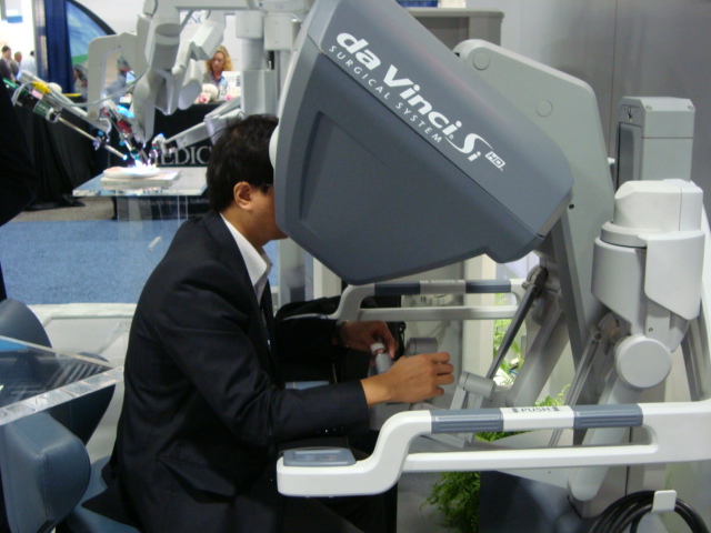
da Vinci Robotic system at the 2012 Academy meeting
The da Vinci Surgical System comprises three components: a surgeon’s console, a patient-side robotic cart with 4 arms manipulated by the surgeon (one to control the camera and three to manipulate instruments), and a high-definition 3D vision system. Articulating surgical instruments are mounted on the robotic arms which are introduced into the body through cannula's. The original telesurgery robotic system that the da Vinci was based on was developed at SRI International in Menlo Park with grant support from DARPA and NASA. Although the telesurgical robot was originally intended to facilitate remotely performed surgery in battlefield and other remote environments, it turned out to be more useful for minimally invasive on-site surgery. The patents for the early prototype were sold to Intuitive Surgical in Mountain View, California.
The da Vinci senses the surgeon’s hand movements and translates them electronically into scaled-down micro-movements to manipulate the tiny proprietary instruments. It also detects and filters out any tremors in the surgeon's hand movements, so that they are not duplicated robotically. The camera used in the system provides a true stereoscopic picture transmitted to a surgeon's console.
Drs. Gregory S. Weinstein and Bert W. O'Malley, Jr. of the University of Pennsylvania School of Medicine's Department of Otorhinolaryngology: Head and Neck Surgery founded the world's first TransOral Robotic Surgery (TORS) program at Penn Medicine in 2004, where they developed and researched the TORS approach for a variety of robotic surgical neck approaches for both malignant and benign tumors of the mouth, voice box, tonsil, tongue and other parts of the throat. TORS has dramatically improved the way head and neck cancers are treated, by achieving complete tumour removal while preserving speech and swallowing. It is a game changer for the way oropharyngeal tumors will be treated in the years to come. Head and neck tumor treatments often involve a combination of surgery, radiation therapy, and chemotherapy. In many cases, surgery offers the greatest chance of cure; yet conventional surgery may require an almost ear-to-ear incision across the throat or splitting the jaw, resulting in speech and swallowing deficits for patients. In comparison, the minimally invasive TORS approach, which accesses the surgical site through the mouth, has been shown to improve long term swallowing function and reduce risk of infection while speeding up the recovery time. When compared to traditional surgeries, after their cancers have been removed successfully, patients have been able to begin swallowing on their own sooner and leave the hospital earlier. TORS outcomes are markedly improved when compared to standard chemotherapy, radiation or traditional open surgical approaches for oropharyngeal cancer.
Obstructive sleep apnea (“sleep apnea”) is a disorder in which the soft tissues of the throat collapse on inhalation during sleep, causing disruption in the normal breathing process. Uvulo-palato-pharyngoplasty has been used to treat the palatal and pharyngeal collapse component of OSA. Tackling the tongue base component of the collapse was not always successful. Now TORS has been employed to successfully reduce the tongue base with considerably less morbidity.
(TORS - Tongue base reduction)
Robotic surgery though the mouth is a safe and effective way to remove tumors of the throat and voice box, The preliminary study examined the outcomes of 13 head and neck cancer patients with tumors located in the region of the throat between the base of the tongue and just above the vocal cords, an area known as the supraglottic region. The study found that the use of robot-assisted surgery to remove these tumors through the mouth took about 25 minutes on average, and that blood loss was minimal -- a little more than three teaspoons, or 15.4 milliliters, on average, per patient. No surgical complications were encountered and 11 of the 13 patients could accept an oral diet within 24 hours. If, on the other hand, these tumors are removed by performing open surgery on the neck, the operation can take around 4 hours to perform, require 7 to 10 days of hospitalization on average and require a tracheostomy tube and a stomach tube, the researchers say.
As the rate of thyroid cancer continues to climb, doctors are urging patients to be more cautious about thyroid nodules, a common disorder that is responsible for a small but growing number of thyroid cancer cases. Until recently, the only way to remove nodules and rule out cancer was through surgery that required a five centimeter incision across the front of the neck.Today however, a new option exists that allows surgeons to access the neck through the armpit, allowing for a biopsy of tissue with no visible scar. The procedure, transaxillary robotic thyroid surgery, utilizes 3D cameras and specially designed robotic arms to create a small incision within the armpit, the mechanical arms work just like hands allowing the specialized surgeon to operate remotely with precise control and movements to remove suspicious nodules. The underarm area has fewer nerve endings than the anterior neck area, so there's less pain, no scarring on the neck, and the incision heals faster.
(Robotic thyroidectomy)
Georgia Health Sciences University surgeons successfully performed a robotic submandibular salivary gland excision through the face lift incision in the hairline. A facelift incision and robotics can help surgeons safely remove a portion of a diseased thyroid from some patients without the characteristic neck scar. "It is outpatient, it doesn't require a surgical drain and it has the advantage of no neck scar," said Dr. David Terris, Chairman of the GHSU Department of Otolaryngology-Head and Neck Surgery. The goal was a no-neck-scar technique that's as safe as conventional thyroidectomy, which involves an incision at the base of the neck to gain immediate access to the thyroid, the surgeons report in The Laryngoscope. The benefits for robotic thyroid surgery include shorter recovery period, less pain in neck following surgery and better preservation of the laryngeal nerves and the parathyroid glands.
Doctors at AIIMS claimed to have performed the first robotic surgery for throat cancer in India, in May 2011. The doctors from AIIMS had been to University of Pennsylvania Hospital, Philadephia, for training in TORS. Similar surgeries were performed at Manipal hospital, Bangalore.
Source:
1)Science Daily
2) Wikipedia
3) Penn Medicine, Dept of Otorhinolaryngology Head & Neck surgey website 4) U tube
The chip is small enough to fit in the cavity of the middle ear. (Credit: Patrick P. Mercier)
In a recent issue of the journal Nature Biotechnology, a team of researchers from MIT, the Massachusetts Eye and Ear Infirmary (MEEI) and the Harvard-MIT Division of Health Sciences and Technology (HST) demonstrated for the first time that cochlear potentials could power implantable electronic devices without impairing hearing. This has far reaching potential in the areas of diagnostics, and other implantable devices like cochlear implants and fully implantable hearing aids. Today the cochlear implants is powered by an external power source. Konstantina Stankovic, an otologic surgeon at MEEI, and HST graduate student Andrew Lysaght implanted electrodes into the cochlea of guinea pigs' ears. Attached to the electrodes were low-power electronic devices developed by MIT's Microsystems Technology Laboratories (MTL). After the implantation, the guinea pigs responded normally to hearing tests, and the devices were able to wirelessly transmit data about the chemical conditions of the ear to an external receiver. . The cochleais the auditory portion of the inner ear. The cochlea is a spiralled, hollow, conical chamber of bone.It houses three chambers or scalae within, which are as follows:
the scala media (containing endolymph), or cochlear duct, a region of high potassium ion concentration that the cilia of the hair cells project into.
This ionic gradient generates the endocochlear potentials. Endocochlear potential (EP) is a battery-like electrochemical gradient found in and actively maintained by the inner ear. The scala vestibuli and scala tympani has a high concentration of Na+ ions, while the scala media higher concentration of K+ ions, The difference in gradients generate this EP. Anantha P Chandrakasan and fellow scientists demonstrated that the mammalian EP can be used as a power source for electronic devices. They achieved this by designing an anatomically sized, ultra-low quiescent-power energy harvester chip integrated with a wireless sensor capable of monitoring the EP itself. The voltage generated by these EP is very low. A device powered by this biological battery can harvest only a small fraction of its power. Low-power chips, however, are precisely the area of expertise of Anantha Chandrakasan's group at MTL. Saurav Bandyopadhyay, a graduate student in Chandrakasan's group, equipped their chip with an ultralow-power radio transmitter: After all, an implantable medical monitor wouldn't be much use if there were no way to retrieve its measurements. The MTL chip also includes power-conversion circuitry in order to amass enough charge to power the radio. In the experiments, the chip itself remained outside the guinea pig's body, but it's small enough to nestle in the cavity of the middle ear. What does it mean for the speciality? This means that we will be able to monitor voltage changes in the endolymph and perilymph on real time basis in patients. This could shed more light in understanding the different stages of Meniere's disease. Imagine being able to know what happens when we perfuse gentamycin or dexamethasone in those patients with Meniere's disease or with Sudden hearing loss. Newer inner ear drugs could be developed in the future using this technology. You could have a fully implantable cochlear implant and a totally implantable hearing aid of a different design in the near future. This technology has tremendous potential for not only otology but even for other branches of modern medicine.
Source:
1)Patrick P Mercier, Andrew C Lysaght, Saurav Bandyopadhyay, Anantha P Chandrakasan, Konstantina M Stankovic. Energy extraction from the biologic battery in the inner ear. Nature Biotechnology, 2012; DOI:10.1038/nbt.2394.
2)Massachusetts Institute of Technology (2012, November 8). Medical devices powered by the ear itself. ScienceDaily.
One of the highlights of the ISCON 2012 conference was the lecture by Prof Ricardo Bento, Sao Paulo, Brazil about his experience with Esteem- the first totally implantable hearing aid. Initially we wondered as to why one would go through the trouble of going through a surgical procedure, instead of just popping a very tiny concealed completely in the canal hearing aid into your ear canal and be done with it. But after hearing Prof. Bento, we came to know as to some people will go through all the trouble. One of the famous implantees is none other than the renowned Otologist, Dr. Michael Glasscock. Prof. Bento showed us the clippings of the procedure and I should say that it is not a easy surgery at all. The FDA took all of five years to approve this implant. He explained how using a conventional canal hearing aid cuts off the resonance created by the external auditory canal, before it strikes the tympanic membrane. It is unique in that it is powered by a battery, which has a life from about 3 to 9 years. This device is also powered by a piezo device which uses the vibration of the tympanic membrane to generate its charge. Since it has no external parts, the user can swim or have a bath and still hear. The device can be controlled with a remote control, and if you take the trouble to turn it off at night, the battery will last even longer. The battery can be changed with a small incision under local anaesthesia. The Esteem® system includes three implantable components (Sound Processor, Sensor, and Driver), and external testing and programming instruments. The Esteem Totally Implantable Hearing System by Envoy Medical Corporation (St. Paul, MN) is a fully implantable system that uses piezoelectric transducers. It uses the eardrum as the microphone. The mechanical signal is detected by a piezoelectric transducer at the head of the incus (the sensor) and converted to an electrical signal that is amplified, filtered, and converted back to a vibratory signal. The processed vibratory signal is then delivered by another piezoelectric transducer (the driver) to the stapes capitulum. The Esteem hearing implant is different from all other microphone-based hearing devices (e.g., hearing aids, other middle ear implants or cochlear implants) because it uses the eardrum to process the incoming sound and thus preserves a natural way of hearing. The Esteem is implanted under the skin behind the ear and in the middle ear space, and therefore invisible. https://www.youtube.com/watch?v=xdBzjR-i9E0 (How the Esteem works- Animation) https://www.youtube.com/watch?v=XdKVaW8krOY (Esteem testimonial-Dr.Glasscock)
The Esteem® device is intended to reduce the effect of moderate to severe hearing loss in patients 18 years of age or older. The Esteem® is used in patients with hearing loss that meet the following criteria:
Stable hearing loss caused by defective hair cells in the inner ear (sensorineural loss) in both ears
Moderate to severe sensorineural hearing loss as defined by Pure Tone Average (PTA) at 500, 100, and 2000 hertz (Hz)
Ability to understand what is heard (speech discrimination test score) greater than or equal to 40% of the words heard during the test.
Normally functioning middle ear (Eustachian) tube
Normal middle ear anatomy
Normal tympanic membrane
Adequate space for the Esteem® implant determined through a high resolution Computed Tomography (CT) scan
Minimum 30 days of experience with appropriately fitted hearing aids.
The Esteem® device should not be used under the following conditions: History of:
post-adolescent chronic middle ear infections
inner ear disorders or recurring vertigo requiring treatment
disorders such as an infection of the mastoid bone (mastoiditis)
swelling in the inner ear (hydrops)
Meniere’s disease (a condition characterized by a ringing sound in the ear(s) or head [tinnitus], hearing loss, vertigo and a feeling of fullness in the ear)
disabling tinnitus which requires treatment
fluctuating air conduction and/or bone conduction hearing loss over the past one year period of 15 decibels (dB) in either direction at two or more hearing frequencies (from 500 to 4000 Hz)
swimmer’s ear (otitis externa) or eczema of the outer ear canal
destructive middle ear disease (cholesteatoma)
central auditory disorders (retrocochlear)
thick scar tissue that continues to grow (keloid formation)
A clinical study of this device involved 60 subjects at 3 investigational centers (57 were implanted with the device). The side-effects observed in the clinical study included:
Taste disturbance: 42% of subjects (14% ongoing after 1 year)
Facial paresis (partial paralysis) / paralysis: 7% of subjects (1% ongoing after 1 year)
Tinnitus: 18% of subjects* (5% ongoing after 1 year) * Some subjects reported having tinnitus prior to the Esteem® implant.
The surgical procedure involved a wide mastoidectomy, a posterior tympanotomy, resection of the long process of the incus, and fixing the driver and the overall system within the middle ear cleft. The tricky part is stripping off the entire mucosa from the stapes superstructure. This is a very delicate maneuver and subluxation of the footplate could be a disaster. This stripping is essential in order to fix the driver to the stapes superstructure using cement. Specific surgical training is needed to accommodate routine surgical steps along with less familiar steps, such as placement of cement material and overall fixation of the system. It is certain that these hearing aids are expensive and requires a lot of expertise in implanting them. It will be preferred b y certain individuals like doctors, musicians and sports persons for whom wearing a conventional hearing aid could be a hindrance in their day to day lives. It will be interesting to see how popular these type of hearing aids will become in the future.